Anatomy Of Back Of Neck - Anatomy Of Back Of Neck Anatomy Drawing Diagram. The majority of neck muscles attach to the hyoid, separating them into two groups: Check spelling or type a new query. It is made up of 24 bones known as vertebrae, according to spine universe. Muscles breathing muscle neck muscles of the neck oral mouth anatomy anterior neck muscles neck muscle anatomy cricoid cricothyroid neck anatomy spine woman. Contains cervical vertebrae and postural muscles.
The respective chapters review in depth all sections of the The neck is connected to the upper back through a series of seven vertebral segments. Hyoid bone explore study unit It runs from the neck to the upper back. From the sides and the back of the neck, the splenius capitis inserts onto the head region, and the splenius.
Upper Cervical Spine Disorders Anatomy Of The Head And Upper Neck from www.spineuniverse.com The suprahyoid and infrahyoid muscles. The occipital bone is a bone that covers the back of your head; The splenius muscles originate at the midline and run laterally and superiorly to their insertions. Contain the common carotid artery, internal. The cervical spine is the top part of the spine. The hard white exterior covering of the tooth is the enamel. Muscle head anatomy vocal organ diagram female neck anatomy neck wireframe head neck human anatomy head artery anatomy face pharynx vector neck degree head anatomy 3d. Contains cervical vertebrae and postural muscles.
The suprahyoid and infrahyoid muscles.
Think of it like a jigsaw puzzle, all the pieces fit in together and are required to get the full picture as to how it works. There are several different layers of muscles in your back. This article gives an overview of the back's structure and its major muscles. The hard white exterior covering of the tooth is the enamel. The splenius muscles originate at the midline and run laterally and superiorly to their insertions. The neck muscles, including the sternocleidomastoid and the trapezius, are responsible for the gross motor movement in the muscular system of the head and neck. Each nerve provides sensation to a specific area of the body called a dermatome. One of the functions of the neck is to act as a conduit for nerves and vessels. Anatomy of the back of your neck. The back muscles stabilize and move the vertebral column, and are grouped according to the lengths and direction of the fascicles. Skin anatomy is often more complex than people think. Muscles make up a large part of the anatomy (structure) of the back. Working in pairs on the left and right sides of the body, these muscles.
The back of the neck is mostly comprised of muscles, as well as the spine. Each nerve provides sensation to a specific area of the body called a dermatome. Back pain is common and might be caused by a problem with a muscle. Anatomy of the back of your neck : The neck is a complex anatomic region between the head and the body.
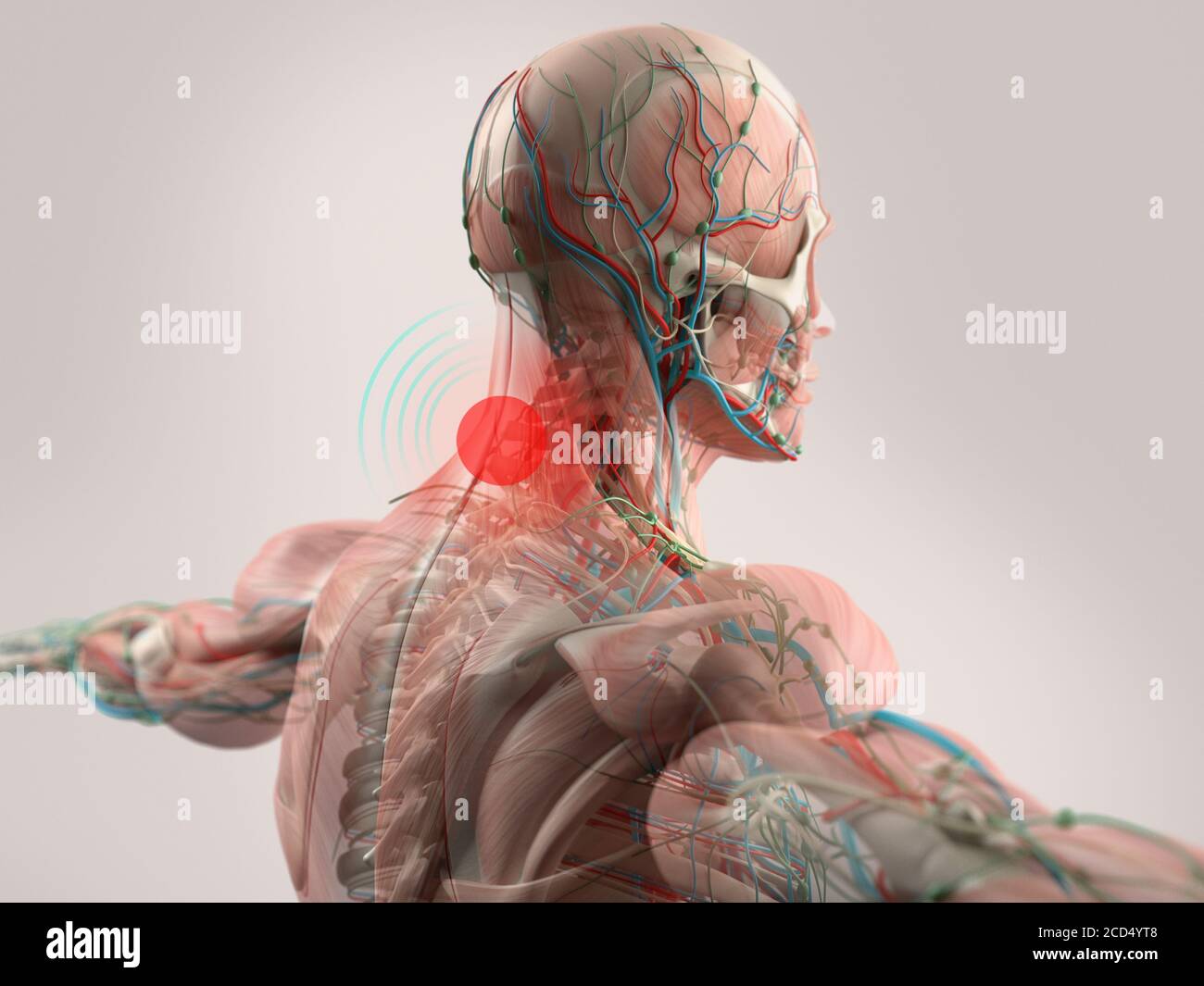
Human Anatomy Neck Pain Head Shoulders And Back Stock Photo Alamy from c8.alamy.com Neck the neck is the start of the spinal column and spinal cord. The hard white exterior covering of the tooth is the enamel. Muscles make up a large part of the anatomy (structure) of the back. Anatomy of the back of your neck : It is made up of 24 bones known as vertebrae, according to spine universe. Below the neck, holding the tooth into the bone, is the root of the tooth. Cervical spine anatomy video the cervical spine has 7 stacked bones called vertebrae, labeled c1 through c7. The cervical spine is the top part of the spine.
As the tooth tapers below the gumline, the neck is formed.
Covering an expanse from the neck to the tailbone, the back muscles are responsible for a broad range of functions, from extending the spine to shrugging the shoulders. Contains cervical vertebrae and postural muscles. Muscle head anatomy vocal organ diagram female neck anatomy neck wireframe head neck human anatomy head artery anatomy face pharynx vector neck degree head anatomy 3d. The cervical spine supports the weight and movement of your head and protects the nerves exiting your brain. Neck anatomy nerves picture there are 8 spinal nerves that originate from the cervical spine. Skin anatomy is often more complex than people think. Neck anatomy explained the neck begins at the base of the skull and connects to the thoracic spine (the upper back). Muscle head anatomy vocal organ diagram female neck anatomy neck wireframe head neck human anatomy head artery anatomy face pharynx vector neck degree head anatomy 3d. Muscles breathing muscle neck muscles of the neck oral mouth anatomy anterior neck muscles neck muscle anatomy cricoid cricothyroid neck anatomy spine woman. Each nerve provides sensation to a specific area of the body called a dermatome. The sternum or breastbone sits in the center of the ribcage. Hyoid bone explore study unit It is made up of bones, discs, muscles, ligaments, nerves and tendons.
The occipital bone is the only bone in your head that connects with your cervical spine (neck). The back muscles stabilize and move the vertebral column, and are grouped according to the lengths and direction of the fascicles. Below the neck, holding the tooth into the bone, is the root of the tooth. Think of it like a jigsaw puzzle, all the pieces fit in together and are required to get the full picture as to how it works. Anatomy of the back of your neck :
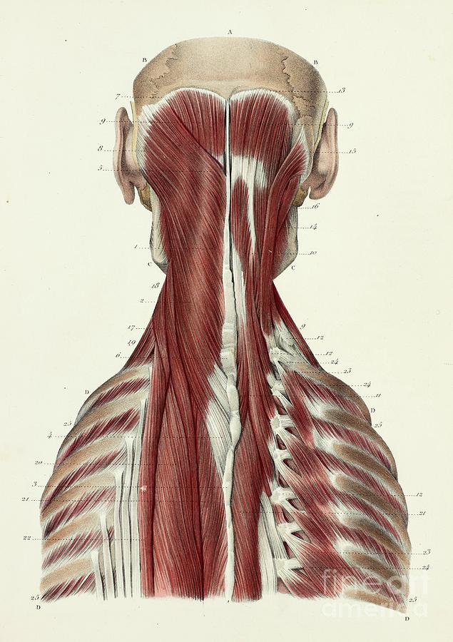
Third Layer Of Back And Neck Muscles Photograph By Science Photo Library from images.fineartamerica.com Covering an expanse from the neck to the tailbone, the back muscles are responsible for a broad range of functions, from extending the spine to shrugging the shoulders. Each nerve provides sensation to a specific area of the body called a dermatome. We did not find results for: It runs from the neck to the upper back. There are several different layers of muscles in your back. Anatomy of the back of your neck. An area called the occiput. This article gives an overview of the back's structure and its major muscles.
They move the head in every direction, pulling the skull and jaw towards the shoulders, spine, and scapula.
It is made up of 24 bones known as vertebrae, according to spine universe. Contains cervical vertebrae and postural muscles. Back of neck anatomy : Neck anatomy explained the neck begins at the base of the skull and connects to the thoracic spine (the upper back). Neck muscles are bodies of tissue that produce motion in the neck when stimulated. The spine is the backbone of the human skeleton. The muscles of your back support your spine, attach your pelvis and shoulders to your trunk, and provide mobility and stability to your trunk and spine. The anatomy of your back muscles can be complex. Check spelling or type a new query. The top of the cervical spine connects to the skull, and the bottom connects to the upper back at about shoulder level. The neck is one of the most complex and intricate structures in our body and includes the spinal cord, which sends messages from the brain to the rest of the body. The splenius muscles originate at the midline and run laterally and superiorly to their insertions. Muscle head anatomy vocal organ diagram female neck anatomy neck wireframe head neck human anatomy head artery anatomy face pharynx vector neck degree head anatomy 3d.